Bacterial Toxins and derivatives
We offer a variety of bacterial toxins, their derivatives, and other proteins that can be used as precision tools for modification of cell metabolic pathways.

Subcategories
-
Bacillus anthracis
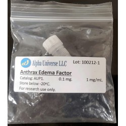
Anthrax toxin is the causative agent of anthrax. It is a three-component exotoxin produced by virulent strains of Bacillus anthracis. Anthrax toxin consists of three nontoxic proteins that associate in binary combinations to form toxic complexes at the surface of mammalian cells. The three proteins of the exotoxin are 83 kDa Protective Antigen (PA), 90 kDa Lethal Factor (LF), and 89 kDa Edema Factor (EF).
Assembly of the toxic complexes begins when PA binds to a cellular receptor and undergoes cleavage by cell protease Furin. As a result, two fragments of PA are generated: an amino-terminal 20-kDa segment of the protein (PA20) that is released, and the carboxyl-terminal 63-kDa fragment (PA63) that remains bound to receptor. Unlike native PA, PA63 forms a ring-shaped heptamers and gains the ability to bind up to three copies of EF and/or LF . Such complexes undergo endocytosis. In the endosomal compartment, the acidic pH causes a conformational change that, eventually, lead to translocation of LF and EF into the cytoplasm.
LF and EF are enzymes. The LF is a Zn2+-dependent protease that cleaves members of the Mitogen-Activated Protein Kinase Kinase family (MAPKK) near to their amino termini. Cleavage of the MAPKKs interferes with MAPK signaling and induces apoptosis. EF is a calmodulin-dependent Adenylate Cyclase. It affects cell homeostasis via catalyzing conversion of ATP into cAMP.
-
Clostridium botulinum
Botulinum neurotoxins are the most potent toxins known to mankind. Seven immunologically distinct serotypes of neurotoxin, designated types A through G (BoNT/A, BoNT/B, BoNT/C, BoNT/D, BoNT/E, BoNT/F, BoNT/G), have been identified.
These homologous toxins specifically target neurons and act through interruption of the process of neurotransmission. This interruption results in muscle paralysis, which, in severe cases of intoxication, leads to death from asphyxiation of humans and animals.
Botulinum neurotoxins are produced by host bacterial cells in the form of single polypeptide chains, which are about 1500 amino acid residues in length (Mr~150 kDa). A single molecule of each toxin possesses three functional domains: receptor-recognizing, transport and catalytic. Catalytic domains of these toxins are Zn2+ metallo-proteases that recognize and selectively cleave proteins involved in targeting of presynaptic vesicles and their fusion with the neuronal plasma membrane. In this way, neurotoxins block neurotransmitter release into the synaptic cleft. Although there is a certain degree of homology between different clostridial neurotoxins, their catalytic domains recognize different substrates: BoNT/B, /D, /F and /G cleave synaptobrevin 2, BoNT/A, /C and /E – synaptosomal-associated protein of 25 kDa (SNAP25) and BoNT/C – syntaxin. Catalytic domains of clostridial neurotoxins are inactive while connected to the rest of the polypeptide chain by a peptide bond and a disulfide bond. As a result of limited proteolysis conducted in the presence of reducing agents, a single polypeptide chain of each neurotoxin can be converted into two chains: light (Mr~50 kDa) and heavy (Mr~100 kDa). The light chain corresponds to the catalytic domain while heavy chains of these neurotoxins contain the receptor-recognizing and transport domains, and are responsible for transport of corresponding light chains into the cytosol of neuronal cells.
-
Clostridium perfringens
Clostridium perfringens is a gram-positive anaerobic bacteria causing numerous gastrointestinal infections in most mammalian species. This microorganism can also cause diseases of skin, subcutaneous, and muscular tissues (gas gangrene or malignant edema). Most, if not all diseases produced by C. perfringens are mediated by one or more of its powerful toxins. Clostridium perfringens can produce up to 16 toxins in various combinations, including:
Alpha toxin is a 43 kDa protein. It contains two domains, an N-terminal domain which is a zinc-dependent phospholipase C, and a C-terminal domain which is involved in membrane binding. Alpha-toxin is a classic example of a toxin that modifies cell membranes by enzymatic activity. It degrades phosphatidylcholine and sphingomyelin, both components of eukaryotic cell membranes. Also, alpha-toxin activates several other membrane and internal cell mechanisms that lead to hemolysis.
Perfringolysin O, also called epsilon toxin, is an example of a sulfhydryl-activated toxin that causes cytolysis by forming pores in cholesterol containing host membranes. It is secreted as a prototoxin (32 kDa), which is converted into a fully active toxin (~1000 times more toxic than the prototoxin) when activated by proteases such as trypsin, chymotrypsin, and a metalloproteinase named lambda toxin that is produced by C. perfringens. After binding to target membranes, the protein assembles into a pre-pore complex. A conformation change leads to insertion in the host membrane and formation of an oligomeric pore complex. Cholesterol may be required for binding to host cell membranes, membrane insertion and pore formation. This toxin is the third most potent clostridial toxin after botulinum toxin and tetanus toxin, with a mouse lethal dose of 100 ng/kg.
Iota toxin is a binary toxin. It is formed by two independent protein components that are not covalently linked, one being the binding component (Ib,100 kDa) and the other the enzymatic component (Ia, 45 kDa). Both components are required for biological activity. The iota toxin components are synthesized as inactive proteins and are proteolytically activated by trypsin or chymotrypsin, removing a 20 kDa N-terminal peptide from the binding component (80 kDa for the active form) and 9 to 11 N-terminal residues from the enzymatic component. Ib triggers the internalization of Ia into the cell by receptor-mediated endocytosis. The mature binding component recognizes a specific cell membrane receptor, heptamerizes and mediates the endocytosis of Ia molecules. The Ia possess ADP-ribosylation activity and modifies G-actin at Arg-177 resulting in depolymerization of actin filaments. The final result of this is a change in morphology (rounding); inhibition of migration and activation of leucocytes; inhibition of smooth muscle contraction; and impairment of endocytosis, exocytosis, and cytokinesis.
-
Clostridium tetani
Clostridium tetani produces a potent neurotoxin called tetanus toxin. It is synthesized as a single polypeptide chain with Mr~150 kDa. Later, it undergoes limited proteolysis that generates two fragments: light (Mr~50 kDa) and heavy (Mr~100 kDa) chains. The mature toxin is composed of a heavy and a light chain that are linked via a disulfide bridge. The heavy chain recognizes specific receptors located on neurons and mediates transport of the light chain inside of them. The light chain that corresponds to the N-terminal part of the originally synthesized polypeptide chain is a metalloendoprotease. Inside of neurons, it selectively cleaves synaptobrevin, an integral membrane protein of synaptic vesicles. This cleavage results in inhibition of the release of neurotransmitters from presynaptic nerve endings.
-
Lysteria monocitogenes
-
Pseudomonas aeruginosa
Pseudomonas Exotoxin A (PE) is the most toxic virulence factor of the pathogenic bacterium P. aeruginosa. This toxin is secreted into the extracellular environment. It molecule is formed by a single polypeptide chain that latter is cleaved by mammalian protease furin to form so-called Active and Binding components. 3D structure of PE has three domains: I (composed of Ia and Ib sub-domains), II and III. Domain Ia (residues 1–252) is responsible for cell recognition. It is a highly specific ligand to the LDL/α2-macroglobulin receptor. Domain II (residues 253–364) possess the furin-cleavable motif (aa 274–280, RHRQPRG) and is involved in translocation of the toxin across membranes. Domain Ib (residues 365–404) is still unclear for its function. Domain III (residues 405–613) catalyze the ADP-ribosylation of elongation factor-2. This modification inactivates eEF-2 and the protein biosynthesis of the host cell comes to a standstill. As a consequence, apoptosis is induced and the host cell irreversibly dies.
ExoS is a bifunctional toxin that is injected directly into host cells. It has two distinct functional domains. The N terminus (amino acids 96-233) encodes a GTPase-activating protein (GAP) activity, whereas the C terminus (amino acids 234-453) encodes a factor-activating ExoS-dependent ADP-ribosyltransferase (ADPRT) activity. The GAP activity inactivates the Rho GTPases Rho (Rac, and Cdc42), whereas the ADPRT activity modifies a variety of host cell substrates, including Ras, ezrin/radixin/moesin (ERM), Rab5, and cyclophilin A.
-
Bordetella pertussis


